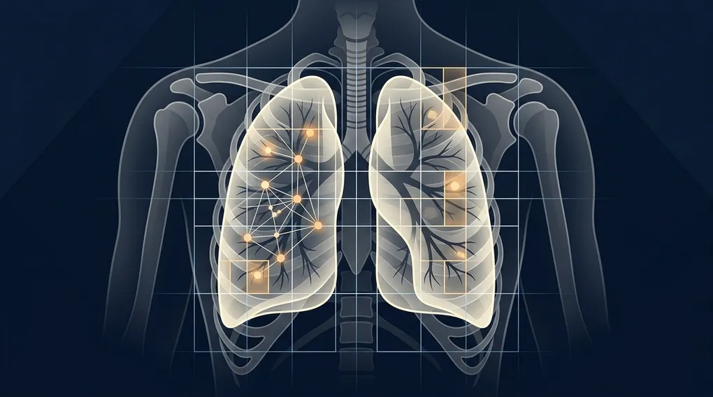
Vision Transformers and CNNs: Hybrid Architectures in Radiological Diagnosis
Table of contents›
In computer-aided diagnosis (Computer-Aided Diagnosis, CAD), machine learning confronts demands qualitatively distinct from conventional computer vision benchmarks. Algorithmic optimization cannot merely seek to maximize global accuracy (Accuracy); it is governed by a fundamental bioethical imperative akin to the classic medical maxim «in dubio pro aegroto» (when in doubt, favor the patient). In a chest radiograph, erroneously classifying an afflicted individual as healthy —incurring a false negative— can delay life-saving treatment for bacterial pneumonia or early alveolar consolidation, with irreversible consequences.
For nearly a decade, deep convolutional neural networks (CNNs) stood as the unquestioned paradigm for medical bioimage analysis. However, the emergence of Vision Transformers (ViT) has fundamentally reshaped our approach to spatial processing. By uniting the complementary inductive strengths of deep convolutions with hierarchical self-attention in hybrid architectures such as CheX-DS, medical artificial intelligence achieves unprecedented sensitivity and diagnostic precision.
From Würzburg to artificial vision: origins of the diagnostic gaze
The human aspiration to scrutinize the interior of the living body without surgical trauma was realized in late nineteenth-century Germany by physicist Wilhelm Conrad Röntgen (1845–1923) (admired for his unblemished experimental rigor and ethical generosity in declining to patent his discovery so that it belonged freely to humanity).
Born in Lennep (Rhenish Prussia) to a modest family of cloth weavers and merchants, Röntgen navigated an irregular educational path: in his youth, he was unjustly expelled from the Utrecht Technical School over a caricature of a teacher that he had not drawn. Undeterred, he passed the competitive entrance examinations for the Swiss Federal Polytechnic in Zurich (ETH), completing his training in mechanical engineering and experimental physics under Rudolf Clausius.
On November 8, 1895, while investigating cathode-ray luminescence in a discharge vacuum tube at the Physikalisches Institut of the University of Würzburg, Röntgen observed an anomaly: a cardboard screen coated with barium platinocyanide crystals emitted a faint green glow even when the discharge tube was completely shielded by thick black cardboard. Röntgen designated this penetrating and previously unrecorded electromagnetic radiation «X-Strahlen» (X-rays, employing the algebraic variable $X$ to signify its unknown nature).
Weeks later, on December 22, 1895, he captured the first anatomical radiograph in history: the hand of his wife, Anna Bertha Ludwig («Hand mit Ringen», hand with rings), in which the dense silhouettes of the phalanges and her wedding band stood out starkly against the soft surrounding tissue.
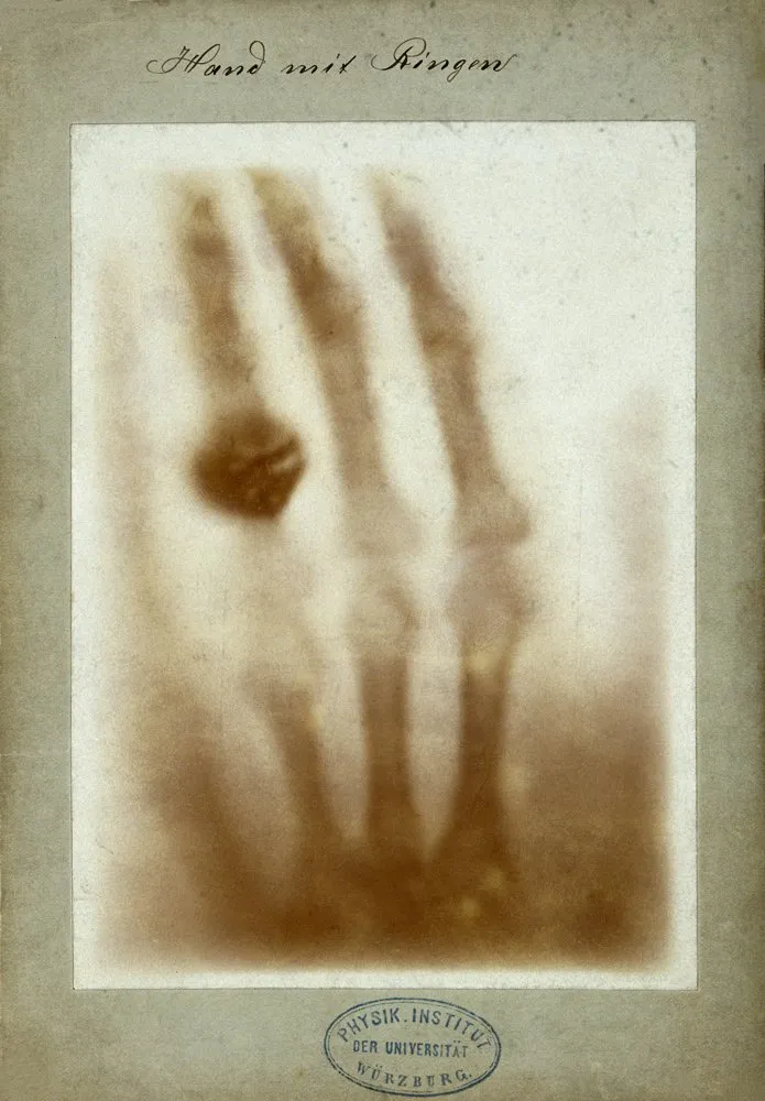 Source: Physikalisches Institut der Universität Würzburg / Wikimedia Commons (Public Domain).
For more than a century, evaluating these radiological densities —black gas, translucent gray soft tissue, and white mineralized bone— relied entirely on the trained clinical eye of the radiologist. With modern computing, the challenge shifted toward designing algorithms capable of providing quantitative, reproducible assistance in reading these subtle anatomical shadows.
The reign of convolution: inductive bias and dense connectivity
The first major breakthrough toward modern automated diagnosis came through deep convolutional neural networks, grounded theoretically by pioneers such as Yann LeCun (admired for his steadfast perseverance during the long years of academic skepticism toward neural networks). Inspired by the receptive fields of mammalian visual cortex described by David Hubel and Torsten Wiesel at Harvard, CNNs introduced a foundational mathematical constraint: the inductive bias (inductive bias) of translational equivariance and invariance.
Through discrete convolution kernels ($K$) with compact spatial support that share weights across the input image $I$:
$$(I * K)(i, j) = \sum_{m} \sum_{n} I(i - m, j - n) K(m, n)$$
CNNs extract hierarchical visual features (tissue boundaries, granular opacities, and interstitial infiltrates) with remarkable parameter efficiency, regardless of where an anomaly is located on the thoracic plane.
However, as networks grew deeper, they encountered the vanishing gradient problem (vanishing gradient problem). To overcome this degradation, Gao Huang and colleagues introduced the DenseNet (Densely Connected Convolutional Networks) architecture in 2017. Rather than relying solely on additive residual skips, every layer $\ell$ within a dense block receives as input the concatenated feature representations from all preceding layers:
$$\mathbf{x}\ell = H\ell([\mathbf{x}_0, \mathbf{x}1, \dots, \mathbf{x}{\ell-1}])$$
where $[\dots]$ denotes concatenation along the channel dimension. In pulmonary pathology, DenseNet121 exhibits outstanding capability: it simultaneously preserves fine spatial details from early layers alongside rich semantic abstractions from deep layers, facilitating the detection of faint infiltrates. Its inherent limitation lies in its receptive horizon: because it processes images via localized sliding windows, it struggles to capture long-range contextual dependencies across distant anatomical structures (such as contralateral pleural effusions or thoracic volume asymmetries).
The self-attention revolution: from Vaswani to the Swin Transformer
In 2017, a research team at Google Brain led by Ashish Vaswani published the landmark paper «Attention Is All You Need», establishing scaled dot-product self-attention (Scaled Dot-Product Attention) for machine translation:
$$\text{Attention}(Q, K, V) = \text{softmax}\left(\frac{QK^T}{\sqrt{d_k}}\right)V$$
where query ($Q$), key ($K$), and value ($V$) projection matrices map data into latent spaces.
Three years later, Alexey Dosovitskiy (admired for his mathematical boldness in toppling the dogma that convolutions were indispensable for computer vision) led the development of the Vision Transformer (ViT) at Google Research. ViT decomposes a two-dimensional image into a linear sequence of non-overlapping patches of fixed size (typically $16 \times 16$ pixels), projecting them as visual tokens analogous to words in natural language.
While ViT excels at modeling global interactions across arbitrary image coordinates, standard self-attention carries a quadratic computational burden:
$$\mathcal{O}(N^2)$$
with respect to the number of patches $N$. In high-resolution clinical radiographs ($1024 \times 1024$ pixels and above), this complexity strains GPU memory limits.
The resolution to this trade-off arrived in 2021 with the Swin Transformer (Shifted Windows), developed by Ze Liu and researchers at Microsoft Research Asia. The Swin Transformer introduces two structural breakthroughs:
- Local window self-attention ($M \times M$): Restricts self-attention computation within local non-overlapping regions of fixed size $M = 7$, yielding linear complexity relative to total patch count: $$\mathcal{O}(4 M^2 N)$$
- Shifted window partitioning (Shifted Window Partitioning): Between consecutive transformer blocks, window boundaries shift by $(\lfloor \frac{M}{2} \rfloor, \lfloor \frac{M}{2} \rfloor)$ pixels, enabling cross-window information exchange and establishing global contextual bridges through a hierarchical feature pyramid.
The CheX-DS architecture: multi-scale synergy and asymmetric formulation
Addressing the clinical intersection between local convolutional precision and broad transformer context, the CheX-DS (Chest X-ray DenseNet & Swin) hybrid architecture was engineered as a synthesis:
   Chest Radiograph (DICOM / Image)
                 │
                 ▼
          ┌───────────────┐
          │  DenseNet121  │  --> Micro-textures & focal opacities
          └───────┬───────┘
                  │ (Enriched intermediate feature maps)
                  ▼
          ┌───────────────┐
          │SwinTransform. │  --> Bilateral context & global dependencies
          └───────┬───────┘
                  │
                  ▼
          ┌───────────────┐
          │Asymmetric Loss│  --> Selective penalization of False Negatives
          └───────┬───────┘
                  │
                  ▼
Audited Clinical Diagnosis (99% Sensitivity / Recall)- Hybrid Information Flow: Enriched feature representations from intermediate stages of DenseNet121 are projected as token embeddings into Swin Transformer stages. This dual-stream design equips the model with both micro-level examination of interstitial patterns and macro-level understanding of thoracic symmetry.
- Weighted Asymmetric Loss (Weighted Asymmetric Loss, ASL): In clinical chest radiography datasets, severe abnormalities are statistically outnumbered by negative cases. Standard cross-entropy is ill-suited because abundant true negatives dampen the learning gradient. CheX-DS implements a specialized asymmetric loss function:
$$\mathcal{L}{\text{asym}} = - (1 - p_m)^{\gamma{+}} y \log(p_m) - \alpha p_m^{\gamma_{-}} (1 - y) \log(1 - p_m)$$
where $y \in {0, 1}$ denotes ground-truth labels, $p$ represents predicted probability, $\gamma_{+}$ and $\gamma_{-}$ are asymmetric focusing exponents, $\alpha$ scales negative penalties, and:
$$p_m = \max(p - m, 0)$$
denotes probability shifted by an asymmetric margin $m \ge 0$. This margin zeroes out gradient updates from easy negatives ($p < m$), concentrating learning capacity on establishing a clean decision boundary for true pathological cases.
Through this training regime, the CheX-DS architecture attained a 99% diagnostic sensitivity (Recall), driving test loss down from $1.03$ (ResNet50 baseline) to $0.29$, ensuring that virtually no pulmonary consolidation escapes automated scrutiny.
From mathematical formulation to auditable interactive deployment
A predictive algorithm offers little real-world utility if confined to a sandbox or operated as an opaque black box. To earn trust in clinical environments, deep learning systems must be interpretable, reproducible, and ergonomic:
- Interpretability via Attention Rollout:
By recursively multiplying attention weight matrices across Swin Transformer layers, the system generates spatial saliency heatmaps directly overlaid on the original radiograph. Clinicians receive not merely a scalar probability score, but an illuminated anatomical map pinpointing the exact tissue regions triggering the diagnostic assessment. - Interactive deployment and containerization:
The inference pipeline is packaged within an interactive Streamlit dashboard, enabling immediate loading of DICOM and high-resolution radiographic images alongside interactive threshold adjustment. The complete environment is containerized via Docker, ensuring deterministic dependency reproducibility across hospital workstations and AWS cloud GPU clusters.
The synergy between the physical nature of X-rays discovered in Würzburg, the mathematical rigor of convolutional inductive biases, and the expressive flexibility of modern Transformers demonstrates that artificial intelligence does not seek to replace clinical judgment, but to augment medical practice with deeper, auditable, and serene tools to preserve human life.
Sources and recommended reading
- The Nobel Prize in Physics 1901: Wilhelm Conrad Röntgen
Official documentation from the Royal Swedish Academy of Sciences on the awarding of the first Nobel Prize in Physics to Wilhelm Röntgen for the discovery of X-rays (X-Strahlen). - arXiv:2010.11929 — An Image is Worth 16x16 Words: Transformers for Image Recognition at Scale (Dosovitskiy et al., 2020)
Foundational paper introducing the Vision Transformer (ViT) by Google Research, establishing pure self-attention on image patches without convolutions. - arXiv:2103.14030 — Swin Transformer: Hierarchical Vision Transformer using Shifted Windows (Liu et al., 2021)
Technical presentation of the Swin Transformer introducing shifted-window partitioning and linear computational complexity. - arXiv:1608.06993 — Densely Connected Convolutional Networks (Huang et al., 2017)
Formal presentation of DenseNet, awarded Best Paper at CVPR 2017 for dense feature reuse. - arXiv:2009.14119 — Asymmetric Loss for Multi-Label Classification (Ben-Baruch et al., 2020)
Mathematical development of asymmetric loss with dynamic margins for extreme class imbalance. - arXiv:1711.05225 — CheXNet: Radiologist-Level Pneumonia Detection on Chest X-Rays with Deep Learning (Rajpurkar et al., 2017)
Stanford Medicine benchmark study applying deep convolutional neural networks to chest radiographs on the NIH ChestX-ray14 dataset.